PAD detection in diabetes: do more, sooner, and smarter
Lower extremity peripheral artery disease (PAD) is common in type 2 diabetes. In fact, people with diabetes are almost twice as likely to develop PAD compared with those without the condition.
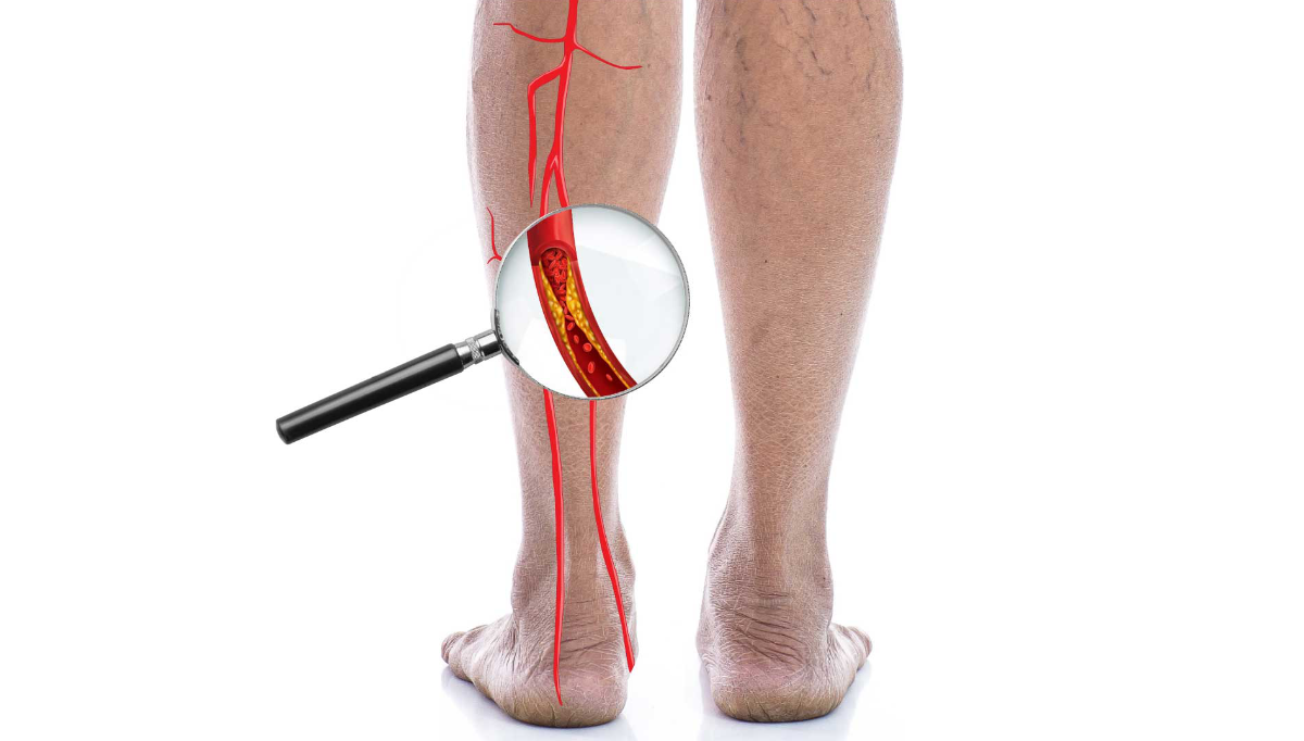
PAD often appears before coronary or cerebrovascular disease, making it an early warning sign of widespread atherosclerosis. Yet, neuropathy often blunts ischemic pain, and medial arterial calcification can make pulses and resting ABI appear deceptively normal. Including objective perfusion assessment as a routine part of diabetes care and taking further steps when the ABI result is unreliable, helps prevent missed diagnoses and ensures timely intervention.
Diabetes accelerates intimal atherosclerosis while promoting medial calcification, resulting in non-compressible arteries and falsely high ABI values. At the same time, sensory neuropathy masks claudication and rest pain, and microvascular dysfunction impairs tissue repair All these conditions combined allow small injuries to progress into ulcers before patients seek help. Nearly half of patients with diabetic foot ulcers also have PAD, and the combination markedly increases the risk of amputation.
Mortality after major amputation remains unacceptably high, with studies showing that up to 40% of patients die within the first year, and nearly 80% within five years. These outcomes underline the importance of early detection and structured vascular evaluation in every diabetic patient’s assessment. In ambulatory care, automated ABI has demonstrated good agreement with Doppler measurements and helps reduce operator dependence, making routine screening both practical and scalable.
Actionable thresholds for vascular assessment
An ankle-brachial index (ABI) of 0.90 or below is an indication of peripheral artery disease (PAD) and should lead to a vascular referral along with immediate measures to protect the feet.
Values in the 0.91–1.00 range are considered borderline. In such cases, the patient’s history and physical findings should be weighed carefully, and a stress ABI may be useful if the patient reports exertional symptoms.
An ABI between 1.00 and 1.40 is typically regarded as normal at rest, but this does not rule out exertional ischemia. If the clinical picture suggests vascular claudication, a stress ABI should be performed.
When the ABI is greater than 1.40, this usually indicates non-compressible arteries. In these cases, the next step is to perform a toe-brachial index (TBI) test, with TBI values of 0.70 or below considered abnormal. A stress ABI is interpreted as positive when the post-exercise ABI decreases by 20% or more, or when the ankle pressure drops by at least 30 mmHg.
From measurement to diagnosis and beyond
Integrating ABI measurement into the structured diabetes management helps make it as routine as checking blood pressure or oxygen saturation. When results are interpreted immediately at the point of care, healthcare professionals can take the next step immediately; whether that means printing the results, offering brief counselling, or arranging additional testing such as TBI or stress ABI, or making a referral when needed.
During the same visit, a comprehensive management approach can be initiated in primary care. This might include guidance on offloading and proper footwear, care for skin and calluses, education on daily foot inspection, support for smoking cessation, and optimization of blood pressure, lipids, and glucose control. Antiplatelet therapy should be prescribed according to clinical guidelines when appropriate.
Recording ABI values in the problem list, setting reminders for follow-up testing, and monitoring referral completion ensure that patients remain connected to the right services. Incorporating ABI into the standard intake template has been shown to increase completion rates: what becomes routine is far more likely to be done.
Cases you may encounter
Non-compressible vessels (ABI >1.40):
This result is common in patients with diabetes or chronic kidney disease. In such cases, the resting ABI is unreliable due to vessel calcification. A toe-brachial index (TBI) should be obtained instead, as toe arteries are less likely to calcify and provide a more accurate reflection of distal perfusion.
Atypical leg symptoms:
Some patients experience tightness, heaviness, or fatigue rather than classic calf pain. When these vascular-sounding symptoms occur alongside an ABI between 0.91 and 1.40, it is better to schedule a stress ABI rather than opt for prolonged observation, as this can help uncover exertional ischemia that may not appear at rest.
Normal ABI, abnormal foot:
A normal ABI does not exclude vascular disease. Any non-healing wound, temperature difference between feet, colour change, or new deformity should prompt vascular evaluation, regardless of the resting ABI result.
When teams embed this approach, decision-making becomes faster and more consistent. Borderline values no longer cause hesitation, exertional symptoms are assessed under exertion, and referrals occur earlier. Clinics begin to see fewer emergency admissions for infected ulcers and greater consistency in preventive foot care. A shared numeric language by using ABI and TBI values with laterality, also strengthens communication and continuity across primary care, diabetology, podiatry, and vascular surgery.
