Pulse wave velocity (PWV) for advanced arterial assessment
Cardiovascular diseases (CVDs) are the leading cause of death globally; the death toll is likely to increase in the future, largely due to population growth and ageing [1]. In 2020 alone, CVDs accounted for approximately 19 million deaths, while deaths caused by cancer, the second leading cause of death, topped at 10 million in the same year [2] [3]. CVDs were traditionally regarded as diseases of affluence, afflicting those from economically developed countries, who could afford the lifestyle causing them. However, the prevalence of CVDs is now rising in low- and middle-income countries [4]. Luckily, there are convenient and cost-effective ways of assessing cardiovascular risk. Pulse wave velocity (PWV) (used to assess arterial stiffness) is an independent predictor of cardiovascular morbidity and mortality [10] [11]; it can be measured in just one minute along with other arterial exams, e.g. together with automated Ankle-Brachial Index (ABI) measurement. All this significantly contributes to advanced arterial assessment.
What is pulse wave velocity (PWV)?
Pulse wave velocity (PWV) is the speed at which the blood pressure pulse propagates through the circulatory system, whether a single artery or a combined length of several arteries [5].
The concept of PWV was introduced by John Crighton Bramwell in the early 20th century; he conducted extensive research on the relationship between the PWV values and arterial wall elasticity [6] [7]. The clinical importance of PWV was recognised in the 1960s, when technology enabled more precise PWV measurements. A significant discovery was that the PWV was higher in diabetic patients and that there was a positive correlation between high PWV and atherosclerotic changes of the arteries [8] [9]. This was expanded further in subsequent research, with strong evidence that PWV is a predictor of future cardiovascular events and all-cause mortality.
Clinical significance of pulse wave velocity
Arterial stiffness as indirectly measured by PWV is an independent predictor of cardiovascular morbidity and mortality [10] [11]. Generally, arterial stiffness increases as a person ages; this is due to the breakdown of the elastin fibers (gradually replaced by stiffer collagen) in the arterial walls, but this process can be accelerated, also due to cardiovascular risk factors (smoking, diabetes, hypertension, obesity, lack of physical activity, etc.) [12]. This makes PWV an integrative biomarker because it reflects the combined effect of modifiable and non-modifiable factors on the arterial health and consequently on the entire cardiovascular system.
According to research, each 1 m/s increase in PWV increases the overall cardiovascular mortality by 12-14% [13]. Also, the 2018 ESC/ESC Guidelines for the Management of Arterial Hypertensionclassify a carotid-femoral PWV of above 10 m/s as asymptomatic organ damage [14]. As a comparison, the mean reference PWV value for healthy individuals under 30 years of age (with optimal or normal blood pressure and no cardiovascular risk factors) is 6.2 m/s [15]. Please note that this reference value is relevant for the European population; the reference values may be different for other populations [16].
Measuring pulse wave velocity (PWV)
There are several methods of measuring PWV. Some are more accurate than others, but are also more expensive and complex to use. The methods can be broadly divided into non-invasive and invasive, and into regional vs. local (by the scope of the assessment). An invasive procedure, usually done quite rarely due to its complexity and costs but regarded as the gold standard for measuring aortic PWV, is catheterisation [17]. Non-invasive methods include magnetic resonance imaging (MRI), which is obviously costly and used only in specific circumstances, and those using pressure cuffs – for measuring carotid-femoral PWV (cfPWV), brachial-ankle PWV (baPWV), and finger-toe PWV (ftPWV) [18] [19] [20]. All have their advantages and limitations; some are more suitable for use in inpatient than outpatient settings, and some are more convenient and cost-effective to use (with acceptable clinical value as an indicator of arterial stiffness). A perfect example of the latter would be the baPWV measurement since it can be done using the same diagnostic devices that can measure the Ankle-Brachial index (ABI).
PWV and ABI can tell a lot about the vascular status of lower extremities (and indirectly about the general state of the cardiovascular system). Something a podiatrist (and many other clinicians) would be interested in.
Pulse wave velocity measurement in podiatry
Podiatrists deal with many diseases and conditions caused by inadequate blood supply. The most prominent example is Peripheral Arterial Disease (PAD), which can result in arterial insufficiency ulcers and ultimately gangrene with associated high mortality rate [21]. PAD is also positively correlated with an increased likelihood of cardiovascular and cerebrovascular events and associated mortality [22], [23]. This is worrisome since the prevalence of PAD is steadily increasing (it is estimated that there were at least 236 million individuals aged 25 years and older with PAD in 2015). Not only is PAD connected with morbidity, but can also limit a person’s mobility and quality of life due to pain while walking (intermittent claudication) or amputations [24] [25]. Moreover, the prevalence and morbidity of PAD can not be effectively calculated based on the number of patients diagnosed because most individuals with PAD are asymptomatic [26].
In addition to aiding in PAD diagnosis and evaluating PAD, the measurement of the Ankle-Brachial Index (ABI) can help differentiate between the two most common types of lower-extremity ulcers: venous ulcers and arterial insufficiency (ischemic) ulcers. The former are the most frequent and represent around 72% of all cases, while the latter are found in between 10% and 30% of patients with lower-extremity ulcers [27]. The rest are neuropathic, lymphatic, and infectious ulcers and ulcers of mixed aetiology, which can be particularly difficult to treat as they often appear in diabetics [27]. Different aetiologies and underlying pathophysiological mechanisms require different treatments [28]. There is significant comorbidity between the two most common types of ulcers, CVDs, PAD and diabetes, itself a risk factor for lower-extremity ulceration as well as PAD and other CVDs [29] [30] [31].
Fortunately, some of the same tools used for diagnosing and assessing PAD can be used for measuring PWV. This gives the podiatrist a more complete picture of the patient’s cardiovascular status and can be helpful when forming a treatment plan. An example is the MESI mTABLET ABI, to which the PWV app can be added.
Solutions like this one put podiatrists in a unique position to identify patients most at risk of future cardiovascular events (even if previously not diagnosed with CVDs like PAD) through the ABI and PWV assessment. The MESI mTABLET diagnostic system also enables immediate ABI and/or PWV result sharing with another specialist.
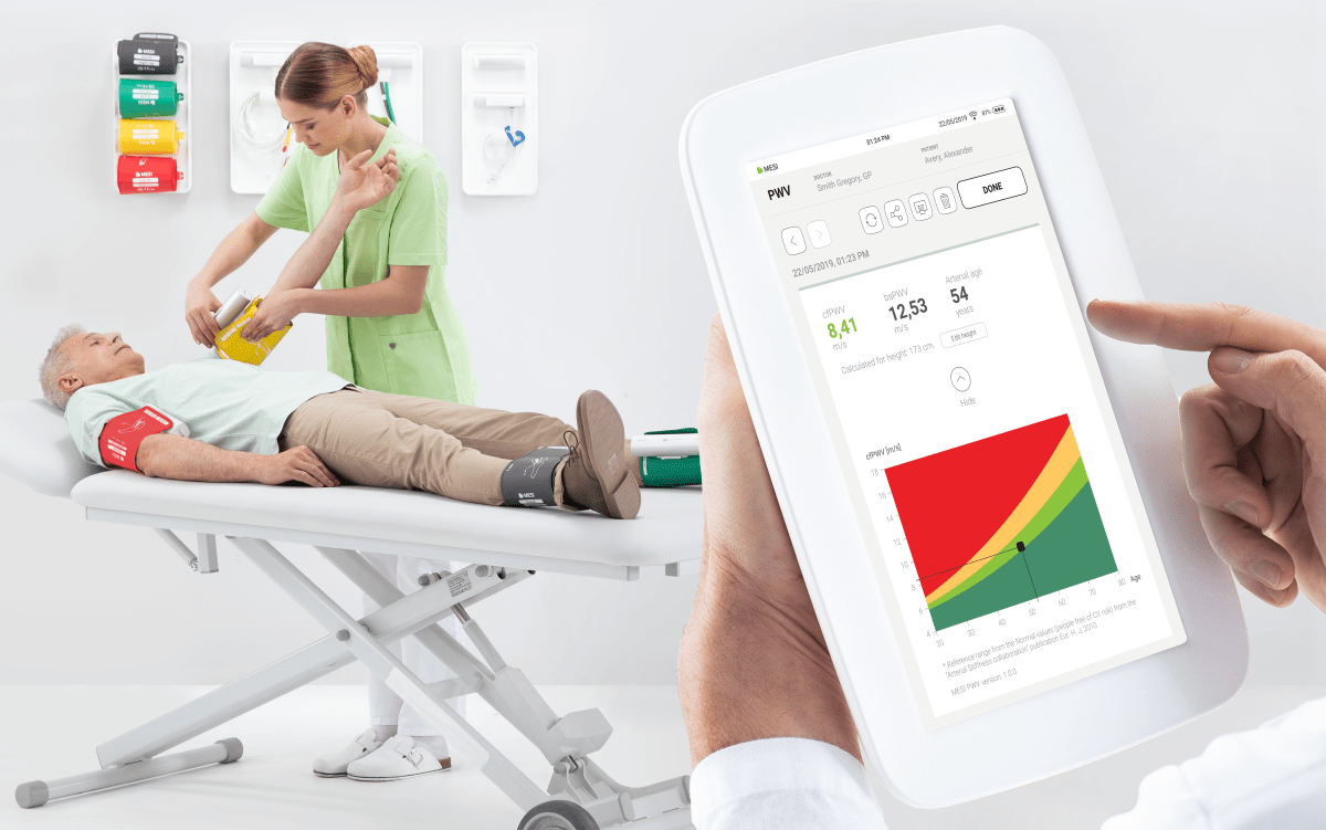
Fast and easy PWV measurement with the PWV app
A prerequisite for measuring the PWV using the MESI mTABLET is the MESI mTABLET ABI module with four inflatable cuffs for measuring ABI and other blood pressure parameters in addition to PWV. The MESI PWV App enables fast and easy, fully automated simultaneous measurement of both the ABI and the PWV (baPWV and cfPWV, calculated using the patient’s height) and the comparison of the latter with reference values (according to the European Arterial Stiffness Collaboration Group). All results are automatically saved in the free MESI mRECORDS cloud-based storage, which can be accessed from every web-enabled device and by anyone with appropriate authorisation. The PWV measurement results can also be instantly shared with other remote specialists and healthcare professionals. The recipient does not need a MESI mTABLET; they receive the result screen in PDF format, with the patient data anonymised according to the applicable legislation.
Find out more about PWVWhat is pulse wave velocity and why is it important in clinical practice? Read more
